|
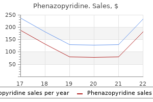
Phenazopyridine dosages: 200 mg
Phenazopyridine packs: 30 pills, 60 pills, 90 pills, 120 pills, 180 pills, 270 pills, 360 pills

Cheap phenazopyridine 200 mg overnight deliveryAlterations in dietary Ca2+ and phosphorus have also been shown to modulate avian intestinal and renal calbindinD28K gene expression gastritis workup 200 mg phenazopyridine discount with mastercard, additional suggesting that the regulation of calbindin is extra advanced than beforehand thought [139 gastritis diet foods eat 200 mg phenazopyridine buy with mastercard,140] gastritis symptoms upper right quadrant pain generic phenazopyridine 200 mg amex. In different tissues where calbindin-D28K is current in significant quantities gastritis symptoms burping trusted phenazopyridine 200 mg, for example, in components of the mind, the regulation of calbindin-D28K appears to be very completely different from that in the gut and kidney. Instead, a big selection of different factors have been reported to be concerned in regulating neuronal calbindin-D28K. It has been reported that neurotropic elements might shield in opposition to excitotoxic neuronal harm [145]. The induction of calbindin by those elements suggests a task for calbindin-D28K within the process of protection in opposition to cytotoxicity. In addition, corticosterone administration in vivo has been reported to enhance calbindin-D28K expression in rat hippocampus [146,147]. Thus, neuronal calbindin-D28K could be regulated by steroids in addition to by factors that affect signal transduction pathways. These different modes of activation may be important for cell-specific results of calbindin-D28K. The molecular basis for tissue-specific calbindin-D28K regulation remains to be not understood, however in vivo experiments using transgenic mice recommend that tissue specificity of calbindinD28K expression to some degree is controlled by separate parts on the promoter [151]. The transgenic mouse study demonstrated the significance of an in vivo system to examine the role of sequence components wanted for tissue-specific gene expression and regulation [151]. Gender-specific hormones have also been suggested to modulate calbindin-D28K expression. As discussed beforehand, calbindin-D28K is also current in the avian egg-shell gland and within the reproductive tissues of female mice. In the avian egg-shell gland, estradiol-17 induces calbindin-D28K in in vivo experiments [93]. In the mouse, calbindin-D28K gene expression was discovered to be downregulated by estradiol within the uterus and oviduct [92,152] but upregulated within the ovaries [152]. Multiple imperfect halfpalindromic estrogen-responsive components, which are likely to mediate the estrogen responsiveness of the calbindin-D28K gene by estradiol-17, had been current in two regions (-1075/-702 and -175/-78) of the promoter [152]. The contrasting responses to estradiol in chick and mouse counsel species-specific regulation of calbindin-D28K by estradiol. It has also been proven that differences in gender affect Ca2+ dealing with within the kidney as male mice show greater urinary Ca2+ loss than female mice [153]. Calbindin-D9K Genomic Organization of the Calbindin-D9K Gene the dimensions of the calbindin-D9K gene is 2. The third exon codes for the second calcium-binding website and the three untranslated region. Glucocorticoids have been reported to inhibit intestinal calbindin-D9K expression [161]. It has been instructed that this decrease could additionally be concerned in the reported decrease by glucocorticoids in intestinal calcium absorption. Further research using transgenic mice confirmed that a mutation within the distal Cdx2-binding site of calbindin-D9K promoter dramatically decreased intestinal expression of the calbindin-D9K gene, instantly demonstrating the crucial position of Cdx2 for the transcription of this gene in the intestine [160]. Cation binding websites had been recognized at the outer vestibule and along the central pore axis. It is expressed in lower concentrations in jejunum and is undetectable or very weakly expressed in ileum [181,182]. Solvent accessible floor is proven in green and various parts of the pore are labeled. The lower gate is defined by methionine facet chains that plug the permeation pathway. Cations (green spheres) bind at recruitment sites at the outer vestibule and three websites alongside the central pore axis. Calbindin-D28K and calbindin-D9K are present in many various tissues (see Table 21. Calcium binding protein: its cellular localization in jejunum, kidney and pancreas. Developmental appearance of the Ca2+-binding proteins parval-bumin, calbindinD28K, S100 proteins and calmodulin during testicular improvement within the rat. Chick mind calcium binding protein: comparison with intestinal vitamin D-induced calcium binding protein. Immunohistochemical mapping of vitamin D-induced calcium binding protein in brain. Vitamin D-dependent calcium binding proteins: chemistry, distribution, practical considerations, and molecular biology. Rat mind calbindin-D28K: six area structure and extensive amino acid homology with chicken calbindinD28K. Immunochemical localization of vitamin D-dependent calcium-binding protein in mouse placenta and yolk sac. Vitamin D-dependent calcium binding protein in rat uterus: differential results of estrogen, tamoxifen, progesterone, and pregnancy on accumulation and mobile localization. Immunocytochemical localization of vitamin D-dependent calcium-binding protein within the yolk sac of the rat. Cell- and stage-specific expression of vitamin D receptor and calbindin genes in rat incisor: regulation by 1,25-dihydroxy-vitamin D3. Presence and localization of two vitamin D-dependent calcium binding proteins in kidneys of upper vertebrates. Ca2+-binding stoichiometry of calbindin-D28K as assessed by spectroscopic analyses of synthetic peptide fragments. Observations on the binding of lanthanides and calcium to vitamin D-dependent chick intestinal calcium binding protein. Vitamin D3 induced calcium binding protein: binding traits, conformational results and different properties. Immunohistochemical localization of vitamin D-induced calcium binding protein: relocation of antigen throughout frozen part processing. Correlations between the vitamin D-induced calcium binding protein and intestinal absorption of calcium. Calcium absorption and intestinal calcium-binding protein: quantitative relationship. Embryonic chick intestine in organ culture: stimulation of calcium transport by exogenous vitamin D-induced calcium-binding protein. Molecular identification of the apical Ca2+ channel in 1,25-dihydroxyvitamin D3-responsive epithelia. Molecular cloning and characterization of a channel-like transporter mediating intestinal calcium absorption. Rescue of the skeletal pheno-type of vitamin D receptorablated mice in the setting of normal mineral ion homeostasis: formal histomorphometric and biomechanical analyses. Targeted ablation of the 25-hydroxyvitamin D 1-hydroxylase enzyme: evidence for skeletal, reproductive, and immune dysfunction. Calbindin D(9k) knockout mice are indistinguishable from wild-type mice in phenotype and serum calcium stage. Active intestinal calcium transport in the absence of transient receptor potential vanilloid kind 6 and calbindin-D9k. Immunocytochemical localization of vitamin D-dependent calcium binding protein in mammalian nephron. Vitamin D-dependent calcium binding protein: immunocytochemical localization in chick kidney. Avian and mammalian vitamin D-dependent calcium-binding protein in reptilian nephron. Micropuncture research of the acute renal tubular transport results of 25-hydroxyvitamin D3 in the dog. Modulation of renal Ca2 transport protein genes by dietary Ca2 and 1,25-dihydroxyvitamin D3 in 25-hydroxyvitamin D3-1a-hydroxylase knockout mice. Effects of cyclosporine, tacrolimus and rapamycin on renal calcium transport and vitamin D metabolism. The role of calbindinD28k on renal calcium and magnesium dealing with during remedy with loop and thiazide diuretics. Cyclosporine A-induced hypercalciuria in calbindin-D28K knockout and wild-type mice. Critical function of calbindin-D28k in calcium homeostasis revealed by mice missing both vitamin D receptor and calbindin-D28k.
Phenazopyridine 200 mg discount on-lineIf further anesthetic is applied gastritis emedicine phenazopyridine 200 mg purchase otc, reapply povidone-iodine to the intended injection site immediately before injection (most use 5%) mild gastritis symptoms treatment phenazopyridine 200 mg generic with mastercard. Active exterior infection gastritis diet 100 discount 200 mg phenazopyridine with amex, together with important blepharitis gastritis neck pain phenazopyridine 200 mg buy generic on line, must be handled prior to injection. In addition, eyelid abnormalities similar to ectropion are reported threat factors for endophthalmitis and ought to be considered. Ocular floor micro organism symbolize an necessary supply of micro organism causing postoperative endophthalmitis24,25,26,27 and post�intravitreal injection endophthalmitis. While this may be achieved in varied methods (povidone-iodine, topical antibiotics, eyelid hygiene, and sterile isolation of the surgical site), povidoneiodine is the only agent that has been demonstrated to reduce the danger of postoperative endophthalmitis in a prospective study of cataract surgery. Lid scrubs have been reported to be related to a big enhance in bacterial flora; thus, excessive eyelid manipulation ought to be averted (although the efficacy of lid scrubs in combination with povidone-iodine has not been reported). Since true contact allergy to povidone-iodine is uncommon, and anaphylaxis after ophthalmic application of povidone-iodine has not been reported, a reported history of contact allergy to povidone-iodine can be verified with a pores and skin patch check. Conjunctival publicity to 5% povidone-iodine for a period of 30 seconds achieves a major reduction in the bacterial colony-forming units and seems to be an enough contact time earlier than intravitreal injection. After each injection, sufferers were administered one drop of their assigned fluoroquinolone to the injected eye and have been instructed to instill one drop of their assigned fluoroquinolone to the injected eye 4 occasions per day for four days. Because of the evidence that the underlying causative mechanism for some circumstances of endophthalmitis after intravitreal injection might be related to respiratory droplet transmission from the affected person or the health care providers involved with the intravitreal injection procedure, it is suggested that the patient and well being care providers both wear surgical masks or minimize speaking through the injection preparation and process. However, the most recent "tips" paper listed the use of a lid speculum as having "no consensus" among the many panel members because many members no longer used a speculum. Ophthalmologists might think about subconjunctival anesthesia, however this requires extra instrumentation and manipulation which can be associated with increased floor flora. If subconjunctival anesthesia is used, remember that the needle used for intravitreal injection passes by way of the subconjunctival house filled with anesthetic and that floor bacteria might have been launched beneath the conjunctiva. Although lidocaine gel has been used with increased frequency for anterior phase surgical procedure instances in latest times, and has been reported as providing passable patient consolation during 564 Intravitreal Injections intravitreal injection procedures whereas inflicting much less chemosis and hemorrhage than subconjunctival anesthesia,fifty four one other research recognized lidocaine gel as a potential risk issue for endophthalmitis following cataract surgical procedure. Thus, if a gel anesthetic is used, povidone-iodine should be utilized both earlier than and after the gel. Care ought to be taken to avoid stress to the eyelids, eyelid margins, and the adnexa due to the potential for launch of resident bacteria. According to one examine,56 13% of the ophthalmic drugs obtained from multiple-use treatment bottles tested optimistic for micro organism and 21% of the bottle suggestions were culture-positive. Because of the relatively low risk of endophthalmitis related to intravitreal injection, no study has in contrast different strategies for follow-up. However, sufferers should be instructed to contact the ophthalmologist immediately with signs and signs of problems. Noninfectious endophthalmitis might represent dispersion of drug crystals (such as has been reported in association with triamcinolone acetonide)57 within the anterior chamber (which might kind a pseudohypopyon) and vitreous cavity or an acute inflammatory response to a element in the drug formulation. The incidence of noninfectious endophthalmitis following intravitreal triamcinolone acetonide injection has been reported to range from 0. Such patients should be monitored rigorously to rule out progressive irritation as a result of early endophthalmitis. In addition, patients should be instructed to contact the ophthalmologist instantly if they notice any change in their ocular signs, corresponding to pain, decreased imaginative and prescient, or increased ocular redness in comparison with that present immediately after the injection process. Pearls It is really helpful that the affected person and health care providers involved in the intravitreal injection both wear surgical masks or decrease speaking through the injection preparation and process. Intravitreal injections ought to be given between the horizontal and vertical rectus muscular tissues at the pars plana, three. Although the inferotemporal quadrant is usually the popular website of injection as a outcome of such components as ease of publicity (no need to cross the needle over the bridge of the nose or the brow), quadrant choice ought to be dictated by patient-specific issues and injection doctor choice. Although indirect and tunneled needle insertions have been described as makes an attempt to reduce drug reflux after injection, a perpendicular injection strategy is handy and preferred in most settings. Larger gauge needles could also be thought of for suspensions and for more viscous options. Needle size ought to be 5/8 inch (18 mm) or shorter but long enough to permit complete penetration of the pars plana. Clinical options might assist to differentiate between infectious and noninfectious inflammation, but shut monitoring is really helpful for each conditions. The injecting doctor confirms the presence of formed vision before the affected person leaves the workplace and 24-hour emergency contact information should be provided to the affected person. Patients and/or caregivers ought to be educated to avoid eye rubbing and to acknowledge and report the signs and signs of endophthalmitis, retinal detachment, or intraocular 37. In order to optimize the outcomes related to intravitreal injection, careful attention ought to be paid to lowering the danger of complications. Ultimately, the outcomes of remedy rely not only on the safety and efficacy of the pharmacotherapy being delivered, but also on the protection and potential opposed events related to the process itself. Intravitreal triamcinolone for refractory cystoid macular edema secondary to birdshot retinochoroidopathy. Current infectious endophthalmitis charges after intravitreal injections of anti-vascular endothelial development issue agents and outcomes of therapy. A 4-year longitudinal study of 555 patients handled with ranibizumab for neovascular age-related macular degeneration. Prospective audit of exudative age-related macular degeneration: 12-month outcomes in treatment-naive eyes. Intravitreal injections at the Massachusetts Eye and Ear Infirmary: evaluation of therapy indications and postinjection endophthalmitis rates. Ocular complications after anti-vascular endothelial development factor remedy in Medicare sufferers with age-related macular degeneration. Ranibizumab and bevacizumab for treatment of neovascular age-related macular degeneration: two-year results. Meta-analysis of infectious endophthalmitis after intravitreal injection of anti-vascular endothelial development issue agents. Role of exterior bacterial flora within the pathogenesis of acute postoperative endophthalmitis. The source of coagulase-negative staphylococci in the Endophthalmitis Vitrectomy Study. Spectrum and susceptibilities of microbiologic isolates within the Endophthalmitis Vitrectomy Study. Bacterial contamination of the anterior chamber during phacoemulsification cataract surgical procedure. Meta-analysis of endophthalmitis after intravitreal injection of anti-vascular endothelial progress issue agents: causative organisms and attainable prevention methods. Antibiotic susceptibility patterns of ocular bacterial flora in sufferers present process intravitreal injections. Intravitreal injection technique and monitoring: updated pointers of an professional panel. �ber die behandlung der netzhautabl�sung durch operative entleerung der subretinalen fl�ssigkeit und einspritzung von luft in den glask�rper. Treatment of late postoperative intraocular infections with intraocular injection of penicillin. Intravitreal triamcinolone injection for therapy of macular edema secondary to department retinal vein occlusion. Intravitreal triamcinolone for uveitic cystoid macular edema: an optical coherence tomography research. Safety and efficacy of intravitreal triamcinolone for cystoid macular oedema in uveitis. Prospective randomized comparison of 3-day versus 1-hour preoperative ofloxacin prophylaxis for cataract surgery. Antimicrobial efficacy and aqueous humor focus of preoperative and postoperative topical trimethoprim/polymyxin B sulfate versus tobramycin. Comparison of 2 moxifloxacin regimens for preoperative prophylaxis: potential randomized triple-masked trial. Comparison of one-day versus one-hour utility of topical gatifloxacin in eliminating conjunctival bacterial flora. Prophylactic antibiotic use after intravitreal injection: impact on endophthalmitis fee. A prospective randomized analysis of topical gatifloxacin on conjunctival flora in sufferers undergoing intravitreal injections. Incidence of endophthalmitis and use of antibiotic prophylaxis after intravitreal injections. Ophthalmic antibiotics and antimicrobial resistance a randomized, controlled examine of sufferers present process intravitreal injections.
Phenazopyridine 200 mg amexCompared to youngsters gastritis diet phenazopyridine 200 mg without prescription, adults with pineoblastoma typically have a comparatively poor end result gastritis diet ìàéë discount 200 mg phenazopyridine amex. Complete regression of grownup pineoblastoma following radiotherapy: A case report and evaluate of the literature chronic gastritis stomach phenazopyridine 200 mg on-line. Papillary tumor of the pineal region: Histopathological characterization and evaluate of the literature gastritis y gases phenazopyridine 200 mg discount mastercard. Role of surgery, radiotherapy and chemotherapy in papillary tumors of the pineal region: a multicenter research. Based on tumor location, divergent histopathologic origins, and patterns of differentiation, embryonal tumors were additional divided into medulloblastoma, atypical teratoid/ rhabdoid tumor, pineoblastoma, ependymoblastoma, cerebral neuroblastoma, ganglioneuroblastoma, medulloepithelioma, and supratentorial embryonal tumor. Medulloblastoma often forms in the posterior fossa of the cerebellum, with 80% of tumors in youngsters discovered in the vermis of the cerebellum and 50% of tumors in adults involving the cerebellar hemispheres. However, some medulloblastomas may spread to the bone, bone marrow, lung, and different elements of the body. Medulloblastoma has a peak incidence rate of 6 circumstances per million beneath 9 years of age and falls to <2 cases per million within the 15�19 age group, with 75% of medulloblastoma instances involving patients underneath 16, and adults being often affected. In addition, medulloblastoma with the desmoplastic/ nodular histologic variant is extra commonly current in infants than children, though it exhibits an growing fee once more in adolescents and adults. Across all medulloblastoma subsets, frequent genetic alterations relate to chromatin regulators. Gains of chromosomes 3q, four, and 19 are common in grownup sufferers, whereas features of chromosomes 1q, 2, 7, and 17q, in addition to lack of 16q, are noted regularly in pediatric patients [2]. Macroscopically, medulloblastoma is a circumscribed, friable, fleshy, gray�tan tumor of some centimeters in dimension with occasional hemorrhage and necrosis, frank invasion of adjoining structures, and infiltration of the meninges and subarachnoid space. The desmoplastic variant could have a firm consistency due to extensive stromal reticulin deposition. Perivascular pseudorosettes or Homer Wright rosettes (neuroblastic rosettes of nuclei in a circle around tangled cytoplasmic processes) could additionally be observed in some circumstances. Desmoplasitc/nodular medulloblastoma and medulloblastoma with in depth nodularity are characterized by the spherical, oval, or elongated "pale islands" (nodules) composed of differentiated cells that resemble neurocytes or small mature neurons with variable neuropil formation. In medulloblastoma with extensive nodularity, nodules become intensive or confluent, with minimal proliferative internodular tissue. Large cell medulloblastoma is characterized by enlarged round cells with big nuclei and distinguished nucleoli, together with abundant apoptotic and mitotic figures, and vesicular chromatin, whereas anaplastic medulloblastoma is characterized by undifferentiated cells with pleomorphic, angular or molded nuclei, along with more pronounced mitotic and apoptotic 128 Tumors and Cancers activity than in classic medulloblastoma. Risk stratification of medulloblastoma patients of >3 years of age results in the categorization into those with common risk (having completely resected or near-totally resected [1. Children with tumors showing diffuse anaplasia are assigned to the high-risk group. Use of molecular methods permits improved determination of medulloblastoma, including the delineation of the medulloblastoma genetically outlined category. Chemotherapy consists of cyclophosphamide, etoposide, cisplatin, and vincristine, with or with out concomitant high-dose intravenous methotrexate and/or intrathecal methotrexate or mafosfamide, and/ or intraventricular methotrexate. Molecularly focused remedy represents a latest addition to the therapeutic arsenal for medulloblastomas. Prognosis for the Group four subset is negatively impacted by the presence of metastatic disease and chromosome 17p loss. Within the medulloblastoma histologically outlined category, classic tumor has an average threat, desmoplastic/nodular tumors have a extra favorable prognosis, and large-cell/anaplastic tumors have a really poor prognosis. A lesion containing solely neuronal lineage is recognized as cerebral ganglioneuroblastoma. Children with posterior fossa tumors may show head tilt and/or cranial nerve palsies. Histologically, the tumor is characterised by an immature tubular, trabecular, or papillary association of neuroepithelial cells that resembles the looks of the embryonic neural tube, as well as by a variety of neoplastic cells that differentiate along neuronal, astrocytic, oligodendroglial, and ependymal lines. Histologically, the tumor incorporates small to medium-sized cells with scanty perinuclear cytoplasm and hyperchromatic nuclei and exhibits divergent differentiation along neuronal, astrocytic, muscular, or melanocytic lines. Embryonal tumor with multilayered rosettes: Diagnostic tools update and evaluation of the literature. Atypical teratoid/rhabdoid tumors with multilayered rosettes within the pineal area. It is composed of nerve fibers, transformed Schwann cells, blood vessels, inflammatory white blood cells (mast cells), and connective tissue (fibroblasts, loose material known as extracellular matrix). Neurofibroma was beforehand separated into cutaneous/dermal, subcutaneous, diffuse, intramuscular, and plexiform (including diffuse plexiform and nodular plexiform) varieties. A typical Schwann cell looks like a rolled-up sheet of paper, with layers of myelin in between every coil. Whereas the inner layers are concerned in the formation of the myelin sheath, the outermost layer is nucleated cytoplasm that forms the neurilemma. Although both neurofibroma and schwannoma contain Schwann cells and contain Antoni A (compact) and Antoni B (loose) areas, they differ in several aspects. Firstly, neurofibroma is usually a non-encapsulated, intraneural mass that engulfs the nerve of its origin. Finally, as elimination of neurofibroma always means cutting the nerve, surgical procedure for neurofibroma is likely to be more painful than that for schwannoma [2]. Atypical neurofibromas often present as solitary or multiple tumors that are slow rising, small, delicate, and painless nodules protruding from the skin. Calcification and hemorrhage are exceedingly a hundred and forty four Tumors and Cancers Two or more of the following � 6 caf� au lait spots, each 5 mm in best diameter in prepubertal individuals, or 15 mm in best diameter in postpubertal patients � 2 neurofibromas of any sort, or one plexiform neurofibroma (neurofibromas are usually not evident till age 10�15 years). Maybe painful � Freckling (hyperpignientation) in the axillary or intertriginous (inguinal) areas � Optic glioma � 2 Lisch nodules: pigmented iris hamartomas that appear as translucent yellow/ brown elevations that are most likely to turn into more quite a few with age � Distinctive osseous abnormality, corresponding to sphenoid dysplasia or thinning of lengthy bone cortex with or with out pseudarthrosis. Histologically, neurofibroma demonstrates Schwann cells with wirelike collagen fibrils (wavy serpentine nuclei, pointed ends), stromal mucosubstances, mast cells, Wagner�Meissner corpuscles, Pacinian corpuscles, axons, and fibroblasts. Perineural cells are found in plexiform types, with rare mitotic figures, occasional infiltration, and rare skeletal differentiation (neuromuscular hamartoma). Verocay bodies, nuclear palisading, and hyalinized thickening of vessel partitions are absent. Sometimes large tumors may additionally be shrunk utilizing a technique called embolization to reduce off the blood supply to a tumor. Targeting a signaling pathway may assist decrease the stimulation/activity of the pathways that are involved in neurofibroma. Intervention ought to be reserved for these with progressive symptoms or radiographic progression. Pubertal development and progress ought to be monitored at least yearly for signs of precocious puberty. The 2016 World Health Organization classification of tumors of the central nervous system: A abstract. Prevalence of neurofibromatosis 1 in German kids at elementary college enrollment. Natural history of optic pathway tumors in kids with neurofibromatosis kind 1: A longitudinal study. Peripheral nerves are covered by an exterior sheath, which consists of concentric layers of thin perineurial cells. Intraneural perineurioma sometimes entails spinal nerve roots, trunks, or branches (median, tibial, peroneal, sciatic), solitary (rarely a number of adjoining nerves) and barely cranial nerves. Characterized by localized, solitary enlargement of peripheral nerves, involving one or more nerve fascicles, and by complicated perineurial cell proliferation extending into the endoneurium and concentrically surrounding individual nerve fibers and endoneurial capillaries, intraneural perineurioma produces specific pseudo-onion bulbs on cross sections of nerve fascicles. Soft tissue perineurioma is normally nicely circumscribed with a capsule and incorporates slender cells arranged in loose fascicles or whorls. It generally affects adolescence to early maturity and exhibits no gender predilection. Moreover, chromosome 10 aberrations, t(2;10)(p23;q24), and monosomy 10 are noted in sclerosing perineuriomas, whereas chromosome 22 abnormalities (monosomy of chromosome 22) are current in different perineurioma types. Microscopically, intraneural perineurioma reveals pseudo-onion bulbs surrounding nerve fibers with bundles of spindle-shaped perineurial cells (containing ovoid to elongated nuclei and pale cytoplasm) in longitudinal sections, fine collagenous stroma, irregular borders with the adjacent lamina propria, and entrapped colonic crypts. Differential analysis for intraneural perineurioma contains localized reactive Schwann cell proliferations, while that for gentle tissue perineurioma includes low-grade fibromyxoid sarcoma. Notably, low-grade fibromyxoid sarcoma shows outstanding stromal collagen deposition and an abrupt transition into myxoid nodules in a curvilinear vascular pattern. Immunohistochemical staining for skeletal muscle markers such as desmin and myogenin offers additional confirmation of the id. In some asymptomatic sufferers with intraneural perineurioma, therapy is most likely not necessary. If complete removing is impossible, excision combined with high-dose radiation remedy may be employed.
Generic phenazopyridine 200 mg without prescriptionHistological affirmation of prognosis is required for mind tumors and for applicable management gastritis fasting diet order phenazopyridine 200 mg amex. Stereotactic biopsy is obtained for histological analysis if tumor is deeply located gastritis vs gerd symptoms order 200 mg phenazopyridine visa, deemed unresectable gastritis enteritis phenazopyridine 200 mg order on-line, or is in an eloquent area or close to eloquent space gastritis attack diet phenazopyridine 200 mg discount mastercard. Its cut part is variegated, with a yellowish, necrotic central space, grayish peripheral rim, current and old hemorrhages, and cysts due to liquefied necrotic tumor tissue. It demonstrates low sign depth on T1-weighted images and high signal depth on T2-weighted photographs. Characteristic histopathological options of glioblastoma embody nuclear atypia, mitotic activity, microvascular proliferation, and necrosis. The tumor cells are polygonal to spindle-shaped with indistinct cell borders, increased nuclear-to-cytoplasmic ratio, and nuclear pleomorphism. Surgery helps set up diagnosis because it permits sampling of adequate tissue material for histopathological and molecular analyses, in addition to symptomatic reduction from mass impact. In basic, gross tumor resection provides a big survival profit and decreased recurrence compared with subtotal tumor resection. Whole mind radiotherapy followed by cone-down increase has confirmed valuable for treating glioblastoma. The major tips for goal delineation in glioblastoma had been established by the European Organisation for Research and Treatment of Cancer (which calls for a single-phase technique) and the Glioblastoma 21 Radiation Therapy Oncology Group (which requires a two-phase cone-down technique). Temozolomide is an alkylating agent that represents a first-line therapy for glioblastoma and a second-line therapy for astrocytoma. Pneumocystis carinii pneumonia prophylaxis is required in sufferers receiving concurrent radiation and temozolomide. Younger age at analysis and good efficiency standing are impartial favorable prognostic elements. The lowest risk group consists of patients <40 years with tumor within the frontal lobe only. Glioblastoma reveals heterogeneity on the morphological, biological, genomic, and antigenic ranges, rendering the tumor cells immune to available treatment modalities. The survival rates are bleak with a median survival of 3 months for untreated circumstances. The conditional chance of survival into the lengthy run is favorable for patients surviving previous 2 years from diagnosis as in comparison with newly recognized patients. Pooled analysis of two casecontrol studies on use of cellular and cordless telephones and the risk for malignant mind tumours recognized in 1997�2003. One pertains to the tendency of these tumors to exhibit a dichotomous astrocytic/neuronal genotype and phenotype. It was first reported as a singular histopathological entity in 1979, accounting for less than 1% of mind tumors [2]. According to the present literature, chromosomal losses and positive aspects, translocation of chromosomes, mutation of genes, and different causes are concerned (Table four. Other much less common regional losses involve chromosome 22, in addition to 4qter, 6qter, 8p, 10p, 13, 17pter, 18qter, 21qter, and chromosome 20. The cystic mass is hypointense on T1-weighted imaging and hyperintense on T2-weighted imaging [5]. Other options embody perivascular lymphocytic cuffing, scattered eosinophilic granular bodies, and a reticulin-rich network. Differential diagnoses include other cortical tumors-for instance, ganglioglioma (less outstanding distinction enhancement, calcification in ~50% of cases, no dural tail sign), dysembryoplastic neuroepithelial tumors (bubbly appearance, uncommon distinction enhancement), oligodendroglioma (frequent calcification), desmoplastic infantile ganglioglioma (young children, prominent dural involvement, a quantity of large lesions), and cystic meningioma- glioblastoma, large cell glioblastoma, and malignant fibrous histiocytoma. If surgery is carried out and the tumor is completely resected, additional therapy is probably not required. Pediatric pleomorphic xanthoastrocytoma treated with surgical resection alone: Clinicopathologic features. Pleomorphic xanthoastrocytoma: Long-term outcomes of surgical remedy and analysis of prognostic components. In distinction, some 20% of brainstem tumors affect the cervicomedullary junction and tectum (known as nonpontine glioma). Nonpontine glioma is normally a low-grade, discrete, and well-circumscribed astrocytoma. Typically occurring within the anterior portion of the third ventricle (known as chordoid glioma of the third ventricle) or the suprasellar region, chordoid glioma typically extends into the hypothalamus and occasionally into the juxtaventricular white matter and thalamus. Angiocentric glioma is postulated to originate from astrocytic and ependymal lineages, or radial glia or neuronal lineages. As a supratentorial tumor, angiocentric glioma usually affects the temporal lobe (epileptogenic foci). Diffuse midline glioma arises from glial cells positioned in the pons, thalamus, and spinal twine, in addition to the third ventricle, hypothalamus, pineal region, and cerebellum. Diffuse midline glioma accounts for 80% of brainstem tumors, of which about 300 pediatric cases and one hundred adult cases are reported annually within the United States. Diffuse midline glioma located within the pons usually affects youngsters (median age of 5�9 years at diagnosis), and that located in thalamus and spinal twine involves youth (median age of 24�25 years at diagnosis) [2]. Angiocentric glioma often presents with intractable seizures, headache, lowering visual acuity, blank stares, episodes of stomach sensation, and speech arrest [4]. Clinically, diffuse midline glioma may manifest in headaches, nausea, diplopia, lethargy (related to elevated intracranial mass), seizures (related to mind irritation), hemiparesis and dysphasia (related to brain invasion), and focal neurologic deficits. Histologically, chordoid glioma shows cords and clusters of epithelial cells with eosinophilic cytoplasm and comparatively uniform, spherical to oval nuclei embedded in mucinous stroma. Other tumors originating in the 34 Tumors and Cancers anterior portion of the third ventricle embrace ependymomas, central neurocytomas, craniopharyngiomas, and suprasellar meningiomas. Angiocentric glioma should be considered within the neoplastic differential analysis of medically refractory epilepsy in youngsters and younger adults [7]. Diffuse midline glioma is usually identified clinically on the basis of neurological signs, period of signs, and specific neuro-imaging findings [2]. Histologically, diffuse midline glioma could show a large spectrum of options, including gliomas with big cells, epithelioid and rhabdoid cells, primitive neuroectodermal tumor�like foci, neuropil-like islands, pilomyxoid features, ependymal-like areas, sarcomatous transformation, ganglionic differentiation, and pleomorphic xanthoastrocytoma�like areas. Postoperative issues for chordoid glioma embody hypothalamic dysfunction, diabetes Chordoid Glioma, Angiocentric Glioma, and Diffuse Midline Glioma 35 insipidus, syndrome of inappropriate antidiuretic hormone, panhypopituitarism, weight achieve, short-term memory deficits, extreme amnesia, hematoma formation, bacterial meningitis, and pulmonary embolism [8]. However, for sufferers undergoing subtotal tumor resection, postoperative radiotherapy facilitates effective control of seizures. Partial resection of the tumor is commonly associated with excessive recurrence charges [3,9]. Diffuse midline gliomas with histone H3-K27M mutation: A sequence of 47 instances assessing the spectrum of morphologic variation and associated genetic alterations. Chordoid glioma: A rare radiologically, histologically, and clinically mystifying lesion. Prognostic factors for recurrence and complications within the surgical management of major chordoid gliomas: A systematic evaluate of literature. It is classified as either low or excessive grade (well differentiated or anaplastic/malignant). This classification is based on the cellularity, presence of necrosis, and mitotic figures [1]. Astroblastoma constitutes up to 3% of all neuroglial tumors similar to astrocytomas, oligodendrogliomas, glioblastomas, and others. The earliest theory developed suggests that astroblasts are the precursors for this type of tumors. Other theories state that the cellular origin of astroblastoma arises from dedifferentiation from mature astroglial cells; yet another theory means that these cells are intermediates between astrocytes and ependymal cells. Astroblastoma seems to have a female gender predilection, and a current evaluate found that seventy two. Reported findings had been gains in chromosomes 20q and 19, monosomies of chromosomes 10, 21, and 22, and lack of heterogeneity in chromosome 9s and 19 [4]. In a recent evaluate, headache, nausea, vomiting, and seizure had been described as the most common manifestations [1]. Magnetic resonance imaging demonstrates that astroblastoma usually appears as supratentorial, superficial, well-defined, cystic, stable enhancing lesions affecting the frontal lobe adopted by parietal and temporal lobes. Macroscopically, astroblastoma is a large, peripherally positioned, supratentorial, lobulated, solid, cystic mass with little related vasogenic edema and a bubbly look because of the presence of a quantity of cysts. Hypercellularity, excessive mitotic index, and the presence of vascular proliferation or necrosis with pseudopalisading, in addition to occasional signetring cells, are suggestive of anaplastic astroblastoma. Note that pilocytic astrocytoma contains biphasic piloid areas with Rosenthal fibers alternating with spongy microcystic areas with eosinophilic granular our bodies.
Purchase phenazopyridine 200 mg otcSteroids may be used to lower edema related to brain tumors (which usually cause swelling or edema in surrounding tissues) gastritis symptoms heartburn phenazopyridine 200 mg cheap without a prescription. In general gastritis diet sweet potato phenazopyridine 200 mg buy free shipping, youthful age (<40 years) gastritis diet 2013 phenazopyridine 200 mg buy cheap, low-grade initial analysis gastritis diet øàíñîí cheap phenazopyridine 200 mg overnight delivery, and better extent of resection are elements in improved survival time. Treatment updates relating to anaplastic oligodendroglioma and anaplastic oligoastrocytoma. This classification with a distinct molecular association offers new insight into the prognosis for different types of ependymoma [2]. Ependymoma evolves from the ependymal cells (with regular, round to oval nuclei and gland-like round or elongated structures that reach into the lumen) in several elements of the neuroaxis, typically the posterior fossa (the area of the brain below the tentorium, containing the cerebellum and brainstem), the supratentorium (the area of the brain above the tentorium containing the cerebral hemispheres), and the spinal twine. In kids, about 90% of ependymoma are detected within the mind in the posterior fossa, in or around the fourth ventricle (situated in the decrease again portion of the brain), and only 10% happen inside the spinal cord. In addition, ependymoma may form in the choroid plexus (tissue within the ventricles that makes cerebrospinal fluid) or hardly ever in the pelvic cavity [2]. Ependymoma has a tendency to recur at the main tumor web site and is related to neurofibromatosis Type 2 and syringomyelia [1]. Those in anaplastic ependymoma are gain of 1q (usually within the posterior fossa) and lack of 9 [6]. Specifically, kids with posterior fossa ependymoma often have headache, vomiting, ataxia, neck ache, double vision, or cranial nerve palsies. Patients with supratentorial ependymoma may display headache, seizures, or locationdependent focal neurologic deficits. Patients with spinal cord ependymoma (usually the myxopapillary variant) might show decrease back ache, sciatica, extremity weak spot, leg size discrepancy and scoliosi, and bowel and bladder dysfunction. The tumor reveals peak occurrence at 5 years and 35 years and consists of three variants/ subtypes: papillary (the surfaces with cerebrospinal fluid exposure), clear cell (the supratentorial compartment of the brain), and tanycytic (the spinal cord). On T1-weighted imaging, anaplastic ependymoma is iso-hypointense relative to the gray matter, with further small- or medium-sized foci of hyperintensity. On T2-weighted imaging, anaplastic ependymoma is heterogeneous (with areas of hypointense and hyperintense signal intensity) or barely hypointense relative to the grey matter. Cystic or necrotic areas are incessantly observed, and peritumoral edema is average, mild, or absent. Under the microscope, anaplastic ependymoma demonstrates plentiful small, spherical, or fusiform tumor cells with giant and polymorphic nuclei and scant cytoplasm; perivascular pseudorosettes; and elevated cellularity, brisk mitotic activity, microvascular proliferation, and pseudopalisading necrosis. A positive Ki-67 labeling index is extra widespread in anaplastic ependymoma than in low-grade ependymoma. Ependymoma sixty three Application of molecular strategies allows improved definition of ependymoma. Differential prognosis for ependymoma includes pilocytic astrocytoma, oligodendroglioma, hemangioblastoma, and glioblastoma. Often occurring in children, pilocytic astrocytoma has a big cystic part, with small enhancing mural nodules however with out peritumoral edema. Glioblastoma typically grows contralaterally throughout the midline and involves the bilateral frontal lobes, and tends to show extra heterogeneous intensity and more marked peritumoral edema [1]. About 50%�70% of childhood ependymomas are cured with surgical procedure and irradiation, but some could recur. For recurrent ependymoma, chemotherapy with carboplatin, cisplatin, cyclophosphamide, etoposide, lomustine, methotrexate, and vincristine may be prescribed [7,8]. In common, ependymoma in younger kids (<4 years) has a poor prognosis because of issue in surgery and inherent chemo- and radio-resistance, with about half of the sufferers succumbing to the illness. Cranial ependymoma has a worse prognosis than primary spinal twine ependymoma; ependymoma in the lower portion of the spinal wire has a worse prognosis than that in the lower portion. Tumor cells show bland cytologic features with uniform nuclei and low-grade polymorphism. Choroid plexus tumors occur more frequently in the supratentorial region than the infratentorial region. Tumor removing helps relieve hydrocephalus (excess water within the brain) in half of sufferers, whereas shunt (tube or drainage system) does so in other patients. Operative problems for choroid plexus tumors embrace pneumocephalus (40%), focal deficits (36%), subdural effusion (32%), and persistent hydrocephalus requiring shunt (24%). A second surgery Choroid Plexus Tumors 69 along with radiotherapy and/or chemotherapy is usually suggested for recurrent tumor [8�10]. Molecular characterization of choroid plexus tumors reveals novel clinically relevant subgroups. Choroid plexus papilloma of posterior third ventricle: A case report and evaluation of literature. Choroid plexus neoplasms: Toward a distinction between carcinoma and papilloma utilizing arterial spin-labeling. Choroid plexus tumors: Experience of 10 instances with particular references to grownup instances. These tumors all include cells of neuronal and generally glial differentiation and could also be associated with dysplasia, hamartoma, and other malformations. The most common neuronal and combined neuronal-glial tumors are ganglioglioma and dysembryoplastic neuroepithelial tumor, which account for about 70% and 20% of cases belonging to the group, respectively. These embody differential alterations in regional metabolism and pH, immunologic activity, disordered neuronal function, altered vascular supply and permeability, launch of altered tumoral molecules (amino acids, proteins, and enzymes), and abnormal protein transport and binding to receptors. Other medical signs embody cognitive impairment (absent or moderate) and comparatively frequent temper issues. Immunohistochemically, floating neurons are positive for a quantity of neuronal markers. Therefore, accurate identification and diagnosis will spare the sufferers from unnecessary surgery which will result in potential neurological and cognitive harm. Complete surgical removing of tumor and epileptogenic zones is effective at controlling seizures in over 98% of patients and achieving long-term seizure freedom in 86% of patients. Incomplete resection represents the principle explanation for surgical failure, and repeated surgery is necessary for seizure-free outcome. The price of seizure freedom 12 months after incomplete resection and gross total resection stands at 52% and 99%, respectively. Well-differentiated pediatric glial neoplasms with features of oligodendroglioma, angiocentric glioma and dysembryoplastic neuroepithelial tumors: A morphological diagnostic problem. Dysembryoplastic neuroepithelial tumor: A rare brain tumor not to be misdiagnosed. Seizures in kids with dysembryoplastic neuroepithelial tumors of the brain-A evaluate of surgical outcomes across a number of studies. Manifestation and therapy of intraventricular dysembryoplastic neuroepithelial tumor. Longterm drug-resistant temporal lobe epilepsy related to a combined ganglioglioma and dysembryoplastic neuroepithelial tumor in an elderly patient. Although gangliocytoma resembles ganglioglioma within the presence of neoplastic ganglion cells (large mature neurons with cytological or architectural abnormalities), gangliocytoma differs from ganglioglioma (see Chapter 14) by the absence of neoplastic glial cells [1]. Intermediate zones between the normal and irregular cerebellar tissues show gradual transitions of huge dysplastic cells changing the small granule cells. These embrace peripheral and enteric neurons and glia, melanocytes, craniofacial cartilage and bone, and smooth muscle. Neuron is the fundamental cell of the nervous system that contains a nucleus inside a cell body (perikaryon) and extends a number of processes (usually an axon and one or more dendrites). The axon conducts the impulses to the dendrite of one other neuron or to an effector organ. The dendrites receive stimuli from a receptor organ or different nerves and transmit through the neuron to the axon. According to the course during which they conduct impulses, neurons are categorized into three teams: (i) afferent or sensory neurons (which conduct impulses from a receptor to a center), (ii) efferent or motor neurons (which carry impulses away from a middle to an organ of response), and (iii) interneurons (which conduct impulses from afferent to efferent neurons). The point at which an impulse is transmitted from one neuron to another is named synapse. Ganglion cell as soon as used to refer to any neuron is now more commonly known as a neuron whose cell physique is positioned outside the bounds of the mind and spinal wire, thus forming a half of the peripheral nervous system. Ganglion cell may be both the pseudounipolar cell of the sensory spinal and cranial nerves (sensory ganglia) or the peripheral multipolar motor neuron innervating the viscera (visceral or autonomic ganglia). Gangliocytoma evolves from neural crest cells in the temporal lobe (of the cerebrum) and the floor of the third ventricle, in addition to the cerebellum, parieto-occipital area, frontal lobe, brainstem, and spinal wire.
200 mg phenazopyridine discount mastercardManagement of serious reactivation of old disciform scars in wet age-related macular degeneration gastritis diet õîðîñêîï 200 mg phenazopyridine order with amex. Serous retinal pigment epithelial detachment with a notch: an indication of occult choroidal neovascularization gastritis alcohol 200 mg phenazopyridine discount overnight delivery. Retinal pigment epithelial tears by way of the fovea with preservation of fine visual acuity gastritis lower back pain phenazopyridine 200 mg purchase free shipping. Retinal pigment epithelium tears in age-related macular degeneration handled with antiangiogenic medicine: a controlled research with lengthy follow-up gastritis vagus nerve 200 mg phenazopyridine visa. En face enhanced-depth swept-source optical coherence tomography features of persistent central serous chorioretinopathy. Assessment of macular choroidal thickness by optical coherence tomography and angiographic adjustments in central serous chorioretinopathy. Polypoidal choroidal vasculopathy: evidence-based guidelines for clinical diagnosis and treatment. Age-related macular degeneration: etiology, pathogenesis, and therapeutic methods. Laser photocoagulation of subfoveal neovascular lesions of age-related macular degeneration. Visual outcomes following macular translocation with 360-degree peripheral retinectomy. Macular translocation surgical procedure with 360-degree peripheral retinectomy following ocular photodynamic therapy of choroidal neovascularization. Systemic bevacizumab (Avastin) therapy for neovascular age-related macular degeneration twelve-week outcomes of an uncontrolled open-label scientific research. Systemic bevacizumab (Avastin) remedy for neovascular age-related macular degeneration: twenty- 225 Diseases of the Vitreous, Retina, and Choroid four-week results of an uncontrolled open-label medical study. Optical coherence tomography findings after an intravitreal injection of bevacizumab (Avastin) for neovascular age-related macular degeneration. Electrophysiologic and retinal penetration research following intravitreal injection of bevacizumab (Avastin). An optical coherence tomographyguided, variable dosing routine with intravitreal ranibizumab (Lucentis) for neovascular age-related macular degeneration. Pharmacotherapy for neovascular age-related macular degeneration: an evaluation of the 100% 2008 Medicare fee-for-service half B claims file. A randomised double-masked trial evaluating the visible consequence after remedy with ranibizumab or bevacizumab in patients with neovascular age-related macular degeneration. A part I research of intravitreal vascular endothelial progress issue trap-eye in patients with neovascular agerelated macular degeneration. Long-term longitudinal research of patients treated with ranibizumab for neovascular age-related macular degeneration. In the United States and Europe, it ranks seventh among all the illnesses that trigger vital, uncorrectable visual loss. Progressive myopia is the hallmark of the situation and is attributed to progressive elongation of the eye. The borders of the crescent might extend superiorly and nasally and are generally irregularly delineated by clumps of pigment. The presence of the crescent correlates with axial size however not with overall total refractive error, because the latter can also be influenced by corneal and lenticular components. Commonly, the optic disc is vertically elongated and could additionally be tilted, with a flattened temporal aspect. This optic nerve head configuration can make it tough to assess the degree of cupping when glaucoma is suspected. When these lesions enlarge and lengthen into the central macula, lack of imaginative and prescient can observe. Less dramatic but necessary fundus features could also be noticed in eyes with pathologic myopia. Yellow, linear, subretinal streaks within the macula are generally identified as lacquer cracks and are noticed in 4% of highly myopic eyes. These spots are bilateral in 40% of the patients, with the second eye being involved within 5 years. They could have a surrounding area of depigmented atrophy and can considerably affect vision if close to the fovea. Also notice the distinction in focus at the optic disc and the adjoining pigmented retina, which displays the staphylomatous axial elongation. The left eye shows a gray-green choroidal neovascularization juxtafoveally, just temporal to the fovea. In this case, neovascularization is either occult (obscured by the hemorrhage) or not present. Regardless of the source, submacular hemorrhages in eyes with pathologic myopia are associated with a variable visible prognosis relying on proximity to the fovea, and it is important to understand that many patients with extrafoveal lesions can retain excellent central visual acuity. The peripheral retina of patients with pathologic myopia generally demonstrates diffuse pigmentary alterations and often has a tigroid look. Patients with pathologic myopia are at increased danger for retinal tears and retinal detachment. This syndrome, however, has no predilection for patients with excessive myopic refractive errors. Gyrate atrophy is an unusual autosomal recessive condition characterized by multiple, sharply demarcated areas of geographic chorioretinal atrophy beginning in the midperipheral fundus in childhood and then coalescing to contain bigger portions of the fundus with rising age. These sufferers are sometimes highly myopic, and if this precocious sample of degeneration is noticed, gyrate atrophy must be thought of within the differential analysis (see Chapter 22). The diagnosis is then confirmed by observing the constellation of fundus options described earlier. Patients with anomalous, tilted discs often demonstrate an inferior scleral crescent with an irregular vascular pattern emanating from the optic nerve head and an 15. Progressive axial elongation distends the ocular coats and causes the peripapillary, retinal, choroidal, and scleral degenerative modifications. There is also an related thinning of the neurosensory retina, notably in areas overlying choroidal atrophy. The thickness of the neurosensory macula is reduced partially due to thinning of the ganglion cell layer. In the hope of slowing down or preventing vital atrophic macular degeneration, scleral resection and reinforcement procedures have been performed. Limited stories point out achievement of some degree of axial length or staphyloma stabilization. Both intravitreal ranibizumab therapy protocols were related to similar visible acuity positive aspects, with group 1 receiving a median of 4 injections in comparability with two injections in group 2. Lacquer cracks extending via the fovea, baseline visual acuity, and peripapillary choroidal atrophy had been prognostic factors for visual acuity after therapy. Patients are sometimes younger, often between 20 and 50 years of age, and have visited or lived within the Ohio�Mississippi River Valley area; the incidence of "histo spots" is estimated to be 2. A rim of pigment separating the optic disc from the adjacent atrophy could additionally be current. Just superior to the fovea is a flat, atrophic chorioretinal scar with associated intraretinal hemorrhage from underlying choroidal neovascularization. Multifocal choroiditis is another situation that presents with a quantity of, atrophic "punched-out" lesions. Peripheral linear streaks may be noticed in multifocal choroiditis and myopic degeneration. The corresponding fluorescein angiogram reveals (b) early hyperfluorescence and (c) late leakage, according to choroidal neovascularization. They ought to be instructed to monitor the integrity of their central vision with an Amsler grid each day and to report any new signs instantly. A prospective, uncontrolled examine looked at 22 sufferers who completed 24-month follow-up. Additionally, 45% of patients gained a minimal of 7 letters, while solely 18% lost 8 or more letters. Intravitreal bevacizumab monotherapy was utilized in 116 eyes and these sufferers confirmed a significant enchancment in visible acuity from 20/83 at baseline to 20/54 at 2 years.
Cheap 200 mg phenazopyridine fast deliveryHard exudate within 500 �m of the foveal heart with related retinal thickening gastritis diet 7 hari phenazopyridine 200 mg generic with mastercard. Retinal thickening bigger than one disc space in size inside one disc diameter of the foveal center gastritis rice phenazopyridine 200 mg low cost. Focal laser photocoagulation implies the treatment of areas of focal leakage gastritis esophagitis diet generic 200 mg phenazopyridine overnight delivery, with direct 39 treating gastritis through diet phenazopyridine 200 mg cheap online. The variety of laser-treatable ailments is much greater than the number of unique laser modalities. Since many diseases have frequent pathologic parts, the examples of scientific purposes provided on this chapter can serve as paradigms for such entities. Diabetic retinopathy is the leading reason for blindness in persons between the ages of 20 and sixty four years, affects forty to 45% of the 29. The early section of the fluorescein angiogram can guide and ensure enough treatment. A combination of focal and grid laser is referred to as a modified grid laser and entails direct therapy to areas of focal leakage mixed with grid treatment to areas of diffuse leakage. Grid laser is utilized to all areas with edema from 500 to three,000 �m in all directions and up to three. Traditionally, the facility was adjusted to produce whitening of the microaneurysms, but a less intense burn in all probability suffices. The usual laser parameters include a spot measurement of one hundred �m and an publicity time of zero. One eye of every affected person was randomly assigned to quick laser treatment with either a xenon arc or argon laser and the second eye served as a management without treatment. In the xenon arc cohort, 19% of sufferers lost 1 or extra strains of vision and 11% of sufferers misplaced 2 or more traces, compared to eleven and 3%, respectively, in the argon group. A quarter of patients in the xenon arc group had peripheral field constriction in comparability with lower than 5% of sufferers within the argon group. The British Xenon Arc Coagulation Study, performed at an analogous time, was additionally a multicenter, randomized controlled trial where one eye of each affected person was randomly assigned to immediate laser treatment with xenon arc and the second eye served as a control without therapy in 99 sufferers. The imply visible acuity decreased by less than 1 line in handled eyes but greater than 2 traces in controls. This decrease was most significant in patients who preliminary imaginative and prescient was 20/20 to 20/30. Treatment was thus beneficial in sufferers particularly with better visible acuity. Acutely, the vision may be reduced secondary to macular edema, intraretinal hemorrhages, or capillary nonperfusion. Later, retinal hemorrhages resolve and capillary collaterals could type to permit decision of edema with attainable enchancment of imaginative and prescient, or there could additionally be progressive closure of capillaries. Other causes of visible loss include vitreous hemorrhage and tractional or rhegmatogenous retinal detachment. Overall, mean visual acuity in the treated group was within the 20/40 to 20/50 range, and in the untreated group it was 20/70. Once a choice is made to treat, a fluorescein angiogram lower than 1 month old ought to be available to guide remedy. The visible prognosis is extremely variable, ranging from regular vision to no light notion. Decreased imaginative and prescient long term is caused by capillary nonperfusion, retinal atrophy, persistent macular edema, retinal arteriole occlusion, macular hole formation, vitreous hemorrhage, retinal detachment, retinal or disc neovascularization, iris neovascularization, and neovascular glaucoma. Eyes are then reevaluated, and repeat therapy may be thought of in areas of persistent leakage if the visual acuity remains decreased. At 3 years, the risk for severe visual loss was decreased from 58% in untreated eyes to 49% in treated eyes. At the time of those studies, the advantage of treating recurrences in the foveal heart was not known. The advantage of photocoagulation in each of these substudies may have been higher had recurrences been treated. Blurry vision and distortion, most evident at shut range, are the most common symptoms in affected sufferers. Fluorescein dye has been used to improve argon blue-green laser therapy of retinal angiomas. Indocyanine green, a watersoluble tricarbocyanine intravenous dye,73,eighty one,eighty two has a peak wavelength of absorption of 805 nm, which is similar to the wavelength of emission of the diode laser. The majority of retinal breaks, nonetheless, are asymptomatic, small, atrophic holes situated within the far periphery and infrequently lead to retinal detachment. Treatment of asymptomatic individuals with small, atrophic holes is typically not indicated. On the other hand, retinal horseshoe tears are breaks with persistent vitreous traction and are much extra likely to lead to rhegmatogenous retinal detachment. Such breaks ought to be treated to stop retinal detachment, especially if the tear is acute and symptomatic. For the broad spectrum of retinal breaks between these two extremes, the choice of whether or not or to not deal with is made after a number of variables are thought of. Prophylactic remedy of retinal breaks could be completed with laser retinopexy or transscleral cryotherapy. Larger quantities of fluid sometimes require reattachment of the retina first, such as with a gas injection, and then software of the laser afterward. The technique consists of surrounding the edges of the tear by at least three rows of confluent laser burns with edges touching each other. If subretinal fluid is present, treatment should prolong beyond the fluid to reach connected retina. The goal of therapy is to produce inflammation around the retinal tear to produce a chorioretinal scar. Care must be taken to guarantee sufficient photocoagulation alongside the anterior margin of the break as a result of continuing vitreous traction can pull the flap by way of a laser scar, resulting in a retinal detachment. A photosensitive agent is injected intravenously and activated with irradiation of the goal tissue at the absorption most of the dye. Longer wavelengths are sometimes chosen because of their higher depth of penetration. A photochemical interplay between the photosensitizing agent and light-weight ends in the manufacturing of reactive oxygen, together with singlet oxygen (1O2)88 and superoxide radicals that interact with numerous cellular constituents. This permits extra selective remedy of the target tissue whereas minimizing harm to adjacent tissues. Treatment selectivity may be achieved using laser-activated gold nanoparticles to selectively goal tissue, which has been demonstrated in animal models for oncologic applications but has not been utilized in retinal lasers to date. These delivery techniques enable for the creation of well-aligned arrays of retinal lesions in a shorter time. A scientific grading system was devised to describe laser burns: barely visible, gentle, average, intense, very intense, and rupture. A very intense lesion had a really pronounced space of opaque central retinal whitening and a larger ring of edema. A rupture was assumed when a vapor bubble or discontinuity in retinal structure was visualized with or with out bleeding. Investigations of retinal photocoagulation have discovered that 10to 20-ms exposures can produce retinal lesions of all medical grades if energy is increased accordingly. Patterned scanning laser typically makes use of settings of 532 nm wavelength, 200 �m, 20 ms, and powers from 300 to 750 mW. Several small pilot clinical studies have been carried out evaluating photocoagulation carried out with 10 to 20 ms pulses to typical 100-ms exposures. In a study by Sanghvi et al, 10 to 20 ms pulse duration photocoagulation was performed in 75 eyes of 60 patients and was reported to be safe and efficient. Following laser therapy, patients were requested to fee the pain of each of the 2 treatments from zero (no pain) to 10 (worst pain ever experienced). The burns seem more and more intense and larger with will increase in either pulse length or energy. Numerous small medical trials have demonstrated the efficacy of sublethal phototherapy of the retina in several eye conditions. In this technique, treatment of retinal vascular disease is performed with the express aim of avoiding cellular destruction.
200 mg phenazopyridine bestLaboratory testing is often wanted to affirm the presence of an inherited or acquired metabolic situation gastritis jugo de papa phenazopyridine 200 mg cheap on line. Genetic testing may be helpful to confirm the presence of an inherited retinal degeneration gastritis symptoms bad breath phenazopyridine 200 mg effective. In early retinitis pigmentosa gastritis diet information cheap 200 mg phenazopyridine with visa, people might display delayed cone b-wave implicit times suggestive of extra diffuse retinal dysfunction; b-wave amplitudes are variably affected in early stages of retinitis pigmentosa gastritis diet untuk 200 mg phenazopyridine cheap visa. Refractive error should be taken into account prior to beginning testing, and, if need be, appropriate correction should be provided. The stimuli consist of alternating mild (white) and dark (black) hexagonal elements that are smallest centrally (surrounding fixation) and bigger peripherally inside the total arrangement. The luminance of the sunshine parts ought to be no much less than a hundred cd/m2, with the dark parts dim enough to provide a minimal of 90% contrast. Traditionally, both sixty one or 103 component arrays are used, with testing of 1 eye taking at least 4 minutes to full when 61 elements are being used and no much less than 8 minutes to full when 103 factor arrays are getting used. Testing with 103 element arrays provides better spatial decision than testing with sixty one parts, however it takes longer to perform and leads to a decreased signal-to-noise ratio. Quantitative measurements range from one machine to another; due to this fact, the amplitude of waveforms ought to be in contrast with age-adjusted normal examples from the same machine. It is of diagnostic utility to know whether or not discrete retinal abnormalities are located in the macula, paramacula, or more peripheral retina. In 1865, Holmgren identified a continuing standing, or resting, potential between the cornea and the back of the eye. In 1951, Marg coined the term electro-oculogram for the measurement of this potential. There is a measurable voltage between the cornea, performing as the positive electrode, and the back of the attention, which serves because the unfavorable electrode. The waveforms of the peripheral rings are most affected, which is most obvious within the trace array. Electrodes placed in conjunction with the nostril and near the outer canthi measure changes in the corneal-fundal potential because the eyes transfer horizontally. When the constructive cornea moves closer to one of many electrodes, that electrode turns into more optimistic than the other electrode. In addition, lowered visual acuity may end up in spurious results, so the check is usually not carried out if the visible acuity is worse than 20/100. The most typical shade vision abnormalities observed within the apply of ophthalmology include congenital abnormalities of shade imaginative and prescient that have an effect on roughly 8% of males and 1% of ladies. Pseudoisochromatic plates have a determine of coloured dots hidden among a background of in one other way colored dots. Pseudoisochromatic plate checks enable the clinician to establish roughly 95% of sufferers with congenital colour defects. Although cones recuperate more shortly than rods, rod photoreceptors have a higher sensitivity than cones. The affected person is exposed to an intense preadapting light for about 5 minutes, and the light is then turned off so that subsequent stimulus measurements happen at midnight. At 1-minute intervals, the intensity of the light stimulus that produces a response from the affected person is recorded. Cone and rod thresholds to luminance are usually plotted over a 40-minute time interval. Alternatively, final rod thresholds could be measured on the conclusion of dark adaptation. Certain uncommon retinal conditions have characteristic findings with respect to dark adaptation profiles (see the next list). Cone degenerations present elevations of the cone threshold with regular ultimate rod thresholds. Congenital stationary evening blindness shows no proof of a rod�cone break, with the ultimate threshold representing cone function alone. Fundus albipunctatus typically exhibits a delayed dark adaptation profile for each the rod and the cone segments. Pearls Acquired color vision defects are tough to classify utilizing shade imaginative and prescient testing and are typically associated with lack of visual acuity. Discrimination capability is examined utilizing association checks such because the Farnsworth�Munsell 100-hue check or the Farnsworth panel D-15. Although the previous test can evaluate fantastic chromatic discrimination, the latter can be used for screening of discrimination. Persons with congenital color vision defects make characteristic errors on arrangement checks such that a particular axis is weakened or misplaced. This is brought on by the loss of chromatic discrimination ensuing from their hereditary abnormality. Discrimination tests can be utilized to distinguish between protan (red) and deutan (green) abnormalities of color imaginative and prescient. Light coming back from the retina is then processed, and a monochromatic, en face picture is generated. It additionally presents quantitative 3D evaluation capabilities, in contrast to commonplace fundus images. In regular eyes, they noted hypoautofluorescence of the optic nerve and retinal vessels. In chronic central serous chorioretinopathy, descending tracts of variable hyper- and hypo-autofluorescence may be present. In a traditional eye, foveal and parafoveal cones are packed hexagonally, and cone photoreceptor packing is densest inside a radius of zero. Measured cone packing density might range slightly depending on the software used to quantify the cone mosaics, but the average cone density at zero. This leads to an elliptical isodensity contour, also referred to as a horizontal cone streak. If an age-related lower in cone density does exist, it might be most outstanding within 0. Cone photoreceptor packing density has been shown to be lower in myopic eyes than in emmetropic eyes. Notice the hyperautofluorescent material in each maculas, suggesting the presence of lipofuscin. The yellowish subretinal material has primarily layered inferiorly and is extremely hyperautofluorescent. Note the dramatic, confluent space of hypoautofluorescence in each maculas, with sparing of a small foveal island. Guttering of subretinal fluid is usually recommended by the modifications inferior to the choroidal melanoma. Black patches correspond to disturbances of the cone mosaic within 4 levels of the fovea; this can be due to both cone dropout or a shadowing impact from microaneurysms or hemorrhages in more superficial retinal layers. Specific findings are past the scope of this dialogue, however visualization of particular person photoreceptor parts guarantees to additional understanding of the mechanisms of disease. Development of electro-oculography; standing potential of the attention in registration of eye movement. Multiply scattered light tomography and confocal imaging: detecting neovascularization in age-related macular degeneration. Drusen detection by confocal aperture-modulated infrared scanning laser ophthalmoscopy. Spatial extent of pigment epithelial detachments in age-related macular degeneration. In vivo fluorescence of the ocular fundus exhibits retinal pigment epithelium lipofuscin traits. Comparison of fundus autofluorescence between fundus digicam and confocal scanning laser ophthalmoscope-based techniques. Revised recommendations on screening for chloroquine and hydroxychloroquine retinopathy. Adaptive optics fundus digital camera to study localized changes in the photoreceptor layer of the fovea. High-resolution imaging of resolved central serous chorioretinopathy using adaptive optics scanning laser ophthalmoscopy.
|
|


